This is a place for me to show some of my work, but I will only be posting images that have not been included in a publication here. I encourage you to skim through some of my published work to see what else I have done. My contributions to published articles are clearly indicated in the "Publications" section.
Microscopy

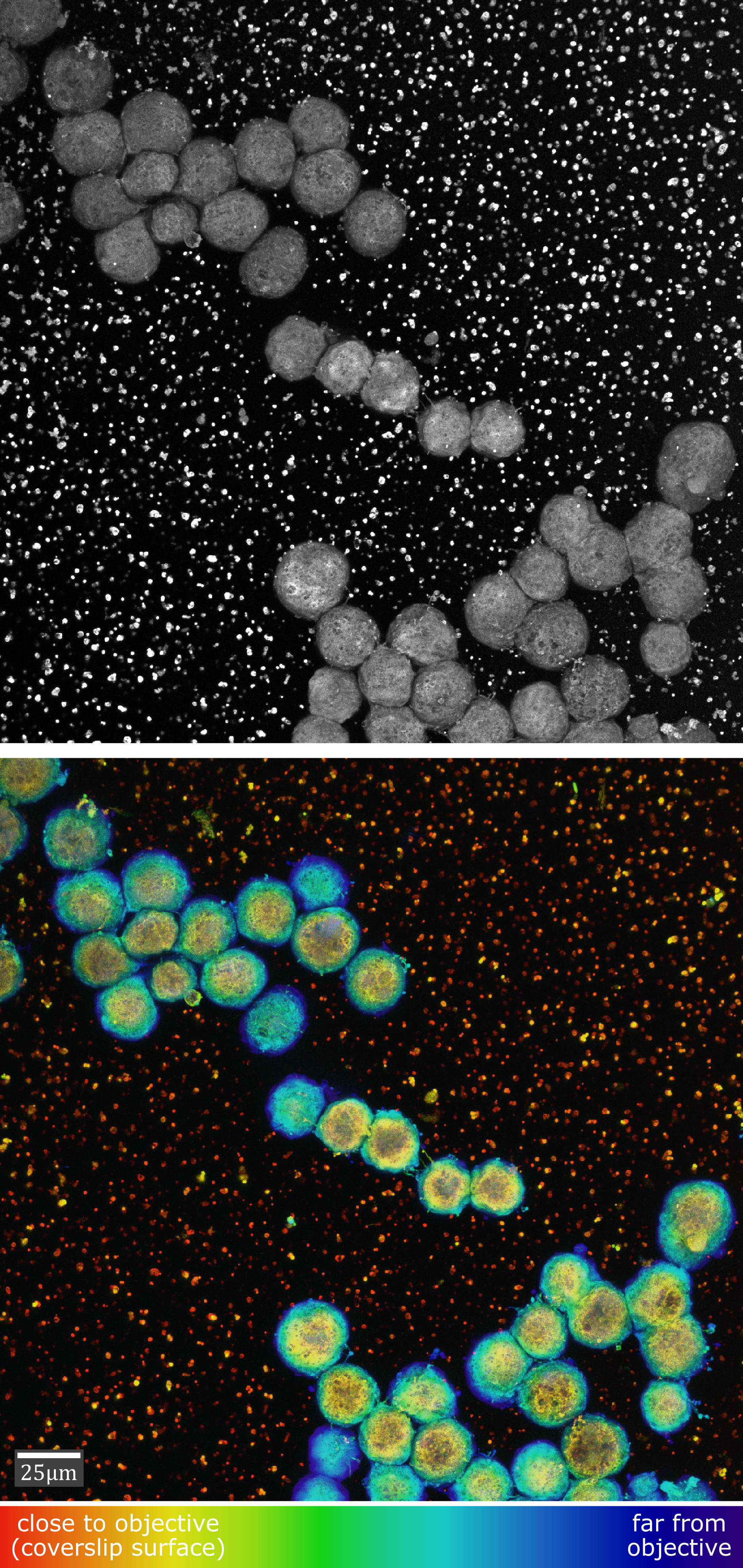
Depth-coded maximum projections of fixed ectoderm cells during reaggregation. Cells are stained with the autofluorescent molecule tannic acid.
Another depth-coded maximum projection of fixed ectoderm cells during reaggregation. Cells are stained with the autofluorescent molecule tannic acid.
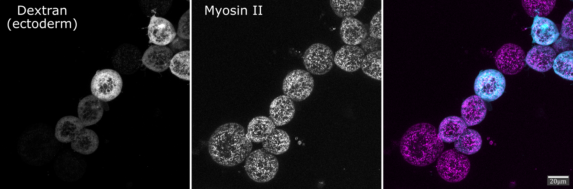
An image of fixed ectoderm and mesoderm cells during reaggregation immunostained for myosin II. Ectoderm cells are shown in cyan, and myosin II in magenta.
Image Analysis
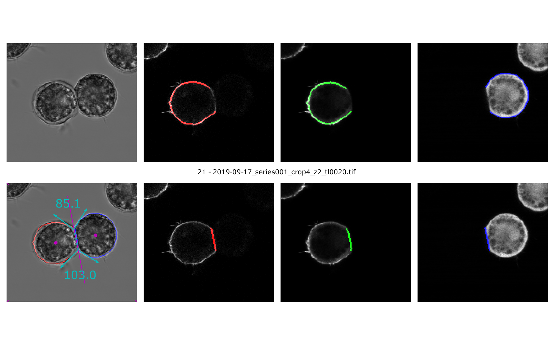
A 2-pixel wide outline of ectoderm cells overlaying a micrograph from the corresponding timepoint.
Data Visualization
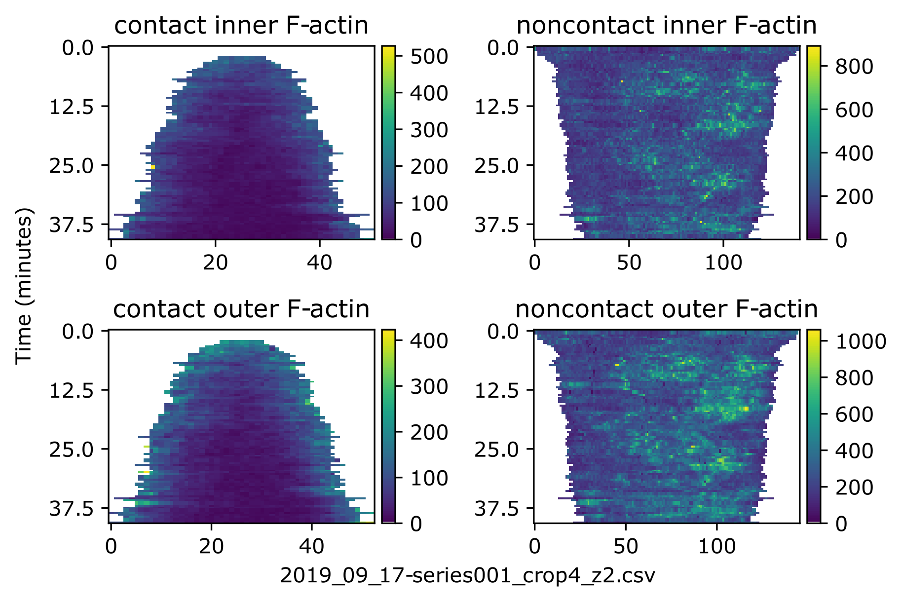
Heatmaps of contact and non-contact F-actin fluorescence.